|
TOTAL EAR CANAL ABLATION (TECA)

TOTAL EAR CANAL ABLATION AND VENTAL BULLA OSTEOTOMY:
THE “TECA” PROCEDURE
|
Sometimes an ear infection is simply hopeless. Perhaps the organism growing is too resistant for treatment. Perhaps the ear canal has actually mineralized from chronic irritation. Perhaps the ear canal is so scarred and narrowed that external cleaning is a useless activity or there is an inflammatory polyp growing into the throat from the middle ear. A tumor may even have emerged in the ear canal. The bottom line is that this degree of irreversible disease requires surgical treatment. In such cases, all the diseased tissue: the entire ear canal, bones of the middle ear etc. are simply removed, the middle ear is drained, and the healthy tissue around the ear is closed. This ends what has generally been a long tribulation of pain, odor, ear cleaning, and expensive veterinary medications and rechecks. The nightmare is over and life is able to go on.
This seems like a beautiful dream to the owner of the pet with end-stage ears but happily it is a realistic dream so long as the process and its associated risks are understood.This surgery essentially removes the ear structures (leaving the ear flap unchanged) and cleans out the tympanic bulla, a structure that most pet owners are unaware of.
Removal of the ear structures is just what it sounds like. The ear canal is cored out and closed up thus removing the entire channel that has been the subject of cleaning and flushing for an undoubtedly long period of time. The area will be closed leaving smooth skin with no opening at the base of the ear flap. The ear flap is basically a satellite dish for sound and, while it most likely has its own dermatitis issues and may continue to have them, the internal ear structures will be gone.
|
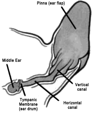
(copyright VIN)
|
As for the tympanic bulla, it must be cracked open and fully cleaned out as it is most likely full of pus, slime, and/or cheesy infectious material. If you are not sure what a tympanic bulla is, you can reach behind your own ear and note that your skull has a smooth round structure. This is your own tympanic bulla. A normal bulla is hollow and air-filled but after years of otitis, it becomes an unreachable, uncleanable source of continued infection. Its lining can even fill with secretory cells that produce more unpleasant material and bone can be destroyed.
Many important nerves travel through the area of the ear and these are exposed for damage in surgery. Damage can be temporary or permanent after surgery. Potential complications will be reviewed in more detail later on.
PREPARING FOR THE TECA
|
1. Radiographs or, even better, CT scanning to assess the tympanic bullae are helpful. Sedation is generally required but it will be useful to know before surgery how bad the bullae look, how narrowed the ear canals are and if they are mineralized, if there is an obvious tumor growing in the area. This helps confirm that this very aggressive surgery is really appropriate for this patient.
2. The ear may be cultured. This helps get the patient on an effective antibiotic right from the beginning. Further cultures may still be required once the bullae are opened.
3. It is important to assess the cranial nerve function of the patient prior to surgery. If these nerves are diseased prior to surgery it is unlikely that they will regain function after surgery. Nerve disease that results from surgery is usually temporary so it is important to know if the nerve problems existed prior to surgery.
The facial nerve runs just near the base of the ear. This nerve controls facial expression. A facial paralysis is not uncommon after long standing ear disease. This means that the patient is slack-jawed on usually one side of the face and may not be able to blink. After a time, the eye usually retracts into the eye socket facilitating tear lubrication so that the loss of blinking does not lead to eye damage. Initially, though, lubricating gels are helpful to the eye.
|
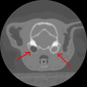
CT Scan from a cat with a polyp in the bulla
shown on the right. The bulla on the left is
normal and is filled with air (as it should be).
Air is black. The fluid and inflammatory
material in the right bulla is gray.
(original graphic by marvistavet.com)
|
Hearing is usually diminished after long term ear infections so further hearing loss after ear ablation may not represent a dramatic change in hearing. Most owners have a good sense of whether or not their pet can hear so it is rarely necessary to formally test hearing. After the ears are ablated, some hearing remains in many patients as sound waves can still be transmitted through the tissues
4. A complete blood panel and urinalysis are important prior to any a major surgical/anesthetic procedure and this procedure is no exception.
WHAT HAPPENS IN SURGERY
|
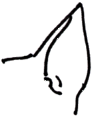
Normal Ear
(original graphic by marvistavet.com)
|

dotted lines indicate
where incision is made
(original graphic by marvistavet.com)
|
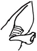
ear canal is then
removed from ear
(original graphic by marvistavet.com)
|

opening into ear is sewn shut
(original graphic by marvistavet.com)
|
|
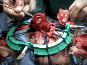
A dog having his ear canal removed (ablated).
The ear canal is the thick cone of tissue being lifted out.
(original graphic by marvistavet.com)
|
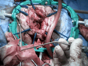
After the ear canal has been removed. A cone-shaped
cavity remains. This will ultimately be closed up.
(original graphic by marvistavet.com)
|
The ears and head are shaved with the patient sleeping and the ear canal is flushed one last time to remove as much infected material as possible. This is done to minimize bacterial contamination of the normal tissue.
The ear canal, both the vertical and horizontal portions, are removed as one long intact curved cylinder. The bones of the middle ear and the ear drum are removed as well.
The bone of the tympanic bulla is exposed and opened. Any material is flushed out and the cellular lining of the bone is scraped away. Any material left inside after closure will lead to chronic drainage of liquid from the area of the incision which is something we do not want. Often an external drain is left in place during the healing period.
Bandaging, oral antibiotics and pain medication can be expected after surgery as can an Elizabethan collar to protect the delicate incisions from scratching.
POTENTIAL COMPLICATIONS
The area of the tympanic bulla and very bottom of the ear canal share space with some important structures. These structures can easily be damaged during surgery or by the inflammation that results during the healing process.
- The “great auricular vasculature” is located on the deep surface of the ear canal being removed. If this vasculature is damaged, the pinna (or furry ear flap) may lose part of its blood supply. Tissue dies along the margin of the ear flap and trimming may be necessary.
- The facial nerve is also located in this vicinity. If the facial nerve is disturbed, a facial paralysis as described above may result. This is one of the more common complications of TECA but is usually temporary. (Facial paralysis after surgery is permanent in 10-15% of cases where it occurs.)
- The “retroglenoid vein” is located just below the tympanic bulla. If this vein is broken, the resulting bleeding is not dangerous but will obscure the visibility of the area making surgery more difficult.
- Sometimes there is enough swelling in the area of the throat to make breathing labored.
- Approximately 5-10% of TECA patients experience chronic drainage from the incision and require a second surgery of some sort to repair the problem. Usually the drainage comes from salivary gland damage, residual cells left in the tympanic bulla, remaining middle ear bone left behind, or the production of more fluid than can drain normally into the throat from the Eustachian tube (the natural connection between ear and throat).
- Hearing is likely to be diminished after surgery though after years of intractable ear disease, hearing is likely diminished already. Hearing should be assumed to be disrupted with this surgery, despite removal of the ear drum. Some dogs are able to hear sounds at normal volumes after the TECA surgery. It is not possible to predict how much more hearing loss an individual will experience after TECA.
CHOLESTEATOMA
The presence of a cholesteatoma can complicate results of the TECA so it is helpful to know in advance if such a structure is present prior to surgery. The cholesteatoma is an expanding structure within the bulla that produces a slimy discharge and destroys tissue including the bones of the bulla as it enlarges. If it is not removed completely (which can be challenging), there will likely be continuing drainage from the area. There may or may not be drainage under the ear evident as early cases frequently manifest with oral pain (especially pain opening the mouth or chewing) and with pain when pressure is applied to the bulla externally. The CT scan, however, should be able to demonstrate bone destruction if a cholesteatoma is present. If a cholesteatoma is suspected, the surgical technique can be appropriately planned.
In most cases, the results are near miraculous.
Patients demonstrate more energy now that their headaches are gone.
There is no more odor, ear cleaning or pain.
This surgery requires advanced skill and referral to a specialist is usually necessary.

Page last updated: 9/2/2024
|